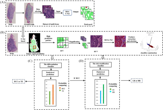
FIGURE 1.

Experimental workflow. (A) Four classes MSA U‐Net based segmentation model (B) Tumor regions identification, nuclear features extraction and selection, and classifier construction. (C) The differentiation classifier generated patch‐level prediction, then compared the probability of BCC and TE, and finally assigned the type with the larger probability. (D) If differentiation classifier predicted the case as BCC, it would transfer to risk stratification classifier for further analysis. BCC, basal cell carcinoma; MSA, multi‐head self‐attention; TE, trichoepithelioma.
