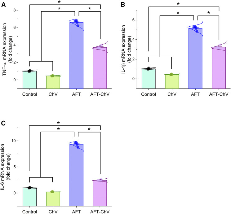
FIGURE 3.
Bar-dot plot panel of mRNA expression of pro-inflammatory cytokines following ChV and/or AFT exposure in the kidney tissue. (A) TNF-α mRNA, (B) IL-1β mRNA, and (C) IL-6 mRNA. AFTs, aflatoxins; ChV, Chlorella vulgaris; IL-1β; interleukin-1β, IL-6, interleukin-6; TNF-α, tumor necrosis factor-α. Values are represented as mean ± SE (*p < 0.05).

