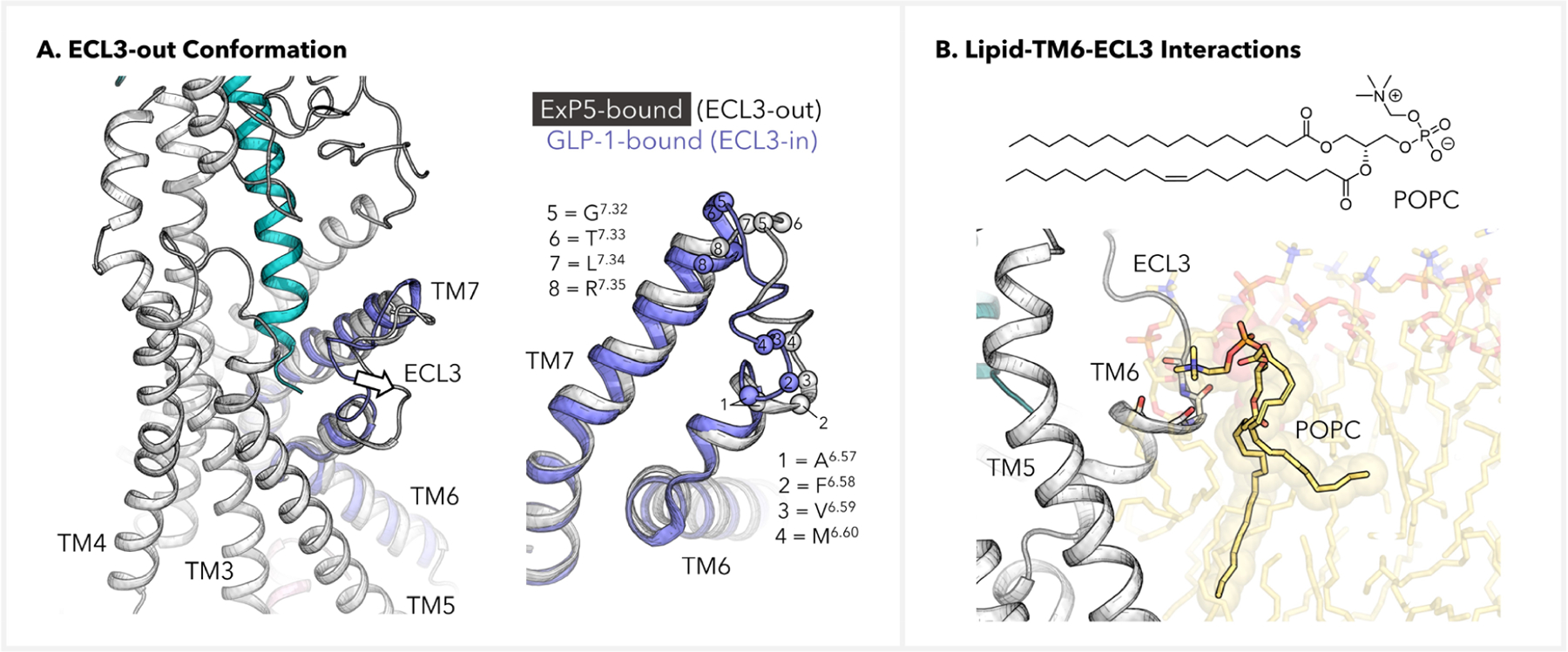
Figure 3.

Lipid-aided induction of the ECL3-out GLP-1R conformation. (A) Depiction of the ECL3-out conformation in the ExP5-bound GLP-1R–Gs complex. Structure of the GLP-1-bound TM6-ECL3-TM7 (blue) was extracted from PDB code 6X18 and was based on alignment of the TMDs. (B) Backbone–POPC interactions at the TM6-ECL3 transition site, where the POPC–NMe3+ group is multianchored to A6.57, F6.58, V6.59, and M6.60. The backbone atoms of these residues are shown as sticks, and the side chains are omitted for clarity.
