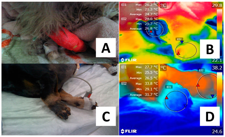
Figure 4.
Difference in the thermal response of patients diagnosed with peripheral vascular alterations. (A) Persian male cat diagnosed with aortic thromboembolism after evaluating clinical signs such as pain on palpation in the right pelvic limb, absence of pulse, cold limb, and mobility difficulty. (B) The phalangeal region of the affected right hindlimb (El1) showed lower temperatures of up to 3.3 °C when compared to the healthy left hindlimb (El2). (C) A four-year-old male Dachshund dog diagnosed with thrombosis due to a secondary liver infection was presented with a reduced perfusion in the left forelimb. Necrosis can be observed. (D) With thermal imaging, it can be observed that the average surface temperature of the phalangeal region of the right forelimb (El2) is 5.2 °C higher than the same region in the affected limb (El1). The explanation for this thermal response is that the presence of a thrombus obstructs blood flow due to occlusion at the arterial level. Thus, the decrease in blood flow has an impact on local heat response. Maximal temperature is indicated with a red triangle and the minimal with a blue triangle. Radiometric images were obtained using a T1020 FLIR thermal camera. Image resolution 1024 × 768; up to 3.1 MP with UltraMax. FLIR Systems, Inc. Wilsonville, OR, USA.

