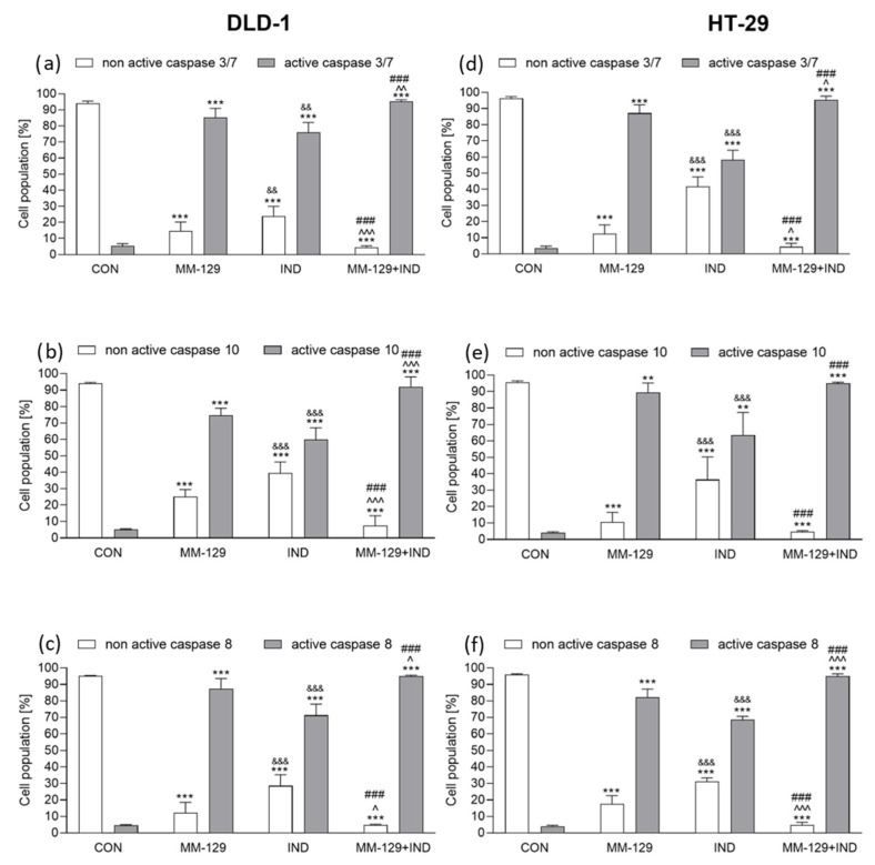
Figure 5.
Flow cytometric analysis of caspase-3/7, caspase-10, and caspase-8 activation in DLD-1 (a–c) and HT-29 (d–f) colon cancer cells exposed to MM-129 (10 µM), IND (200 µM), and MM-129 + IND for 24 h. Values from three independent experiments performed in duplicate were presented as mean ± standard deviation (SD) and analyzed using one-way analysis of variance (ANOVA). ** p < 0.01, *** p < 0.001 vs. CON; ^ p < 0.05, ^^ p < 0.01, ^^^ p < 0.001vs. MM-129; ### p < 0.001 vs. IND; && p < 0.01, &&& p < 0.001 IND vs. MM-129.

