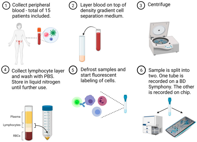
Figure 1.
Overview of experimental study design in a six-step process. Peripheral blood samples were obtained from 15 patients with ovarian cancer. White blood cells were isolated by means of a density gradient centrifugation and frozen until further use. Batches of four to five samples were defrosted and labelled with fluorescent dyes. Each fluorescently stained patient sample was split into two equal parts to perform simultaneous but separate acquisition on a conventional flow cytometer (BD FACSymphony) and our own, silicon, microfluidics-based chip cytometer (Figure created in BioRender).

