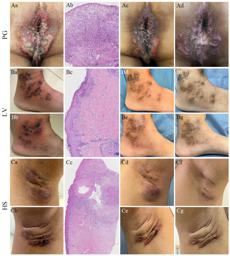
Figure 1.
Patient (A) ulcerative lesions with massive purulent discharges in perianal area before abrocitinib treatment (Aa); extensive infiltration of lymphocytes and neutrophils and necrotising vasculitis (Ab, ×100); significant improvement of skin lesions 1 week after abrocitinib and cyclosporine A treatment (Ac) and 4 weeks after starting abrocitinib treatment (Ad). Patient (B) multiple ulcers and tan necrosis near both ankle joints (Ba, b); cellulose like degeneration of capillary wall, and vascular thrombosis and inflammation (Bc, ×100); markedly improvement of skin lesions 1 week (Bd, e) and 6 weeks (Bf, g) after abrocitinib treatment. Patient (C) nodules and abscesses of the axillary (Ca, b); hyperplasia of the hair follicle infundibulum epithelium and infiltration of lymphocyte (Cc, ×40); improvement of skin lesions 2 weeks after abrocitinib and doxycycline treatment (Cd, e) and 6 weeks after starting abrocitinib treatment (Cf, g).

