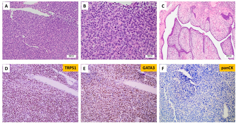
Figure 5.
Malignant phyllodes tumors of the breast with EGFRvIII. (A) Histology shows a poorly differentiated high-grade neoplasm (100×). (B) Analysis under high-power magnification reveals pleomorphic epithelioid tumor cells with a high nuclear-to-cytoplasm ratio and no identifiable definitive epithelial component (200×). (C) A focal area with leaf-like fronds was identified (40×). Tumor cells show diffuse expression of (D) TRPS1 and (E) GATA3 and (F) rare expression of cytokeratin.

