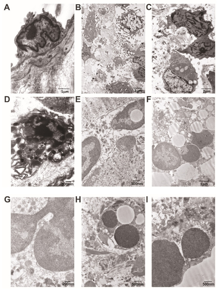
Figure 9.
Representative images obtained using transmission electron microscopy. (A) The endothelium of the valvular leaflet in a Bartonella spp.-caused IE patient is roundly shaped, with few irregular and short microvilli, a wavy nuclear envelope, and marginated chromatin; cytoplasm reveals occasional organelles. (B) A low-power view of the damaged valvular leaflet in a non-Bartonella spp.-caused IE patient. Its content demonstrates various immune cells, including macrophages with well-developed pseudopodia and lipid-laden, are intermixed with collagen microfibrils. (C) In a region nearby, valvular cells reveal a very pronounced and deeply convoluted nuclear envelope; immune cells reveal the extensive expansion of the rough endoplasmic reticulum with content of varying density. (D) An irregularly shaped macrophage with a ruptured plasma membrane reveals a dilated perinuclear space, damaged mitochondria, and some cytoplasmic granules. (E) A neutrophil with a ruptured plasma membrane, wavy-outlined external nuclear membrane, and nuclear inclusion reveals a low-density hyaloplasm containing various granules. Clumps of neutrophil heterochromatin are positioned extracellularly. (F) The free apoptotic nuclei of a neutrophil are surrounded by extracellularly positioned clumps of heterochromatin, cellular debris, and lipid droplets. (G) A high-power view of the neutrophil reveals a nucleus with an irregularly dilated perinuclear space and a ruptured membrane. It is surrounded by a cytoplasm containing granules and autophagic vacuoles. (H) A fragment of the apoptotic neutrophil is enclosed by fibrin, collagen, and cellular debris. (I) A fragment of the neutrophil reveals an irregularly dilated perinuclear space with thread-like material, a ruptured membrane, and various granules positioned both intra- and extracellularly. Scale bars: 1 μm, 2 μm, 2 μm, 500 nm, 500 nm, 1 μm, 200 nm, 500 nm, and 500 nm.

