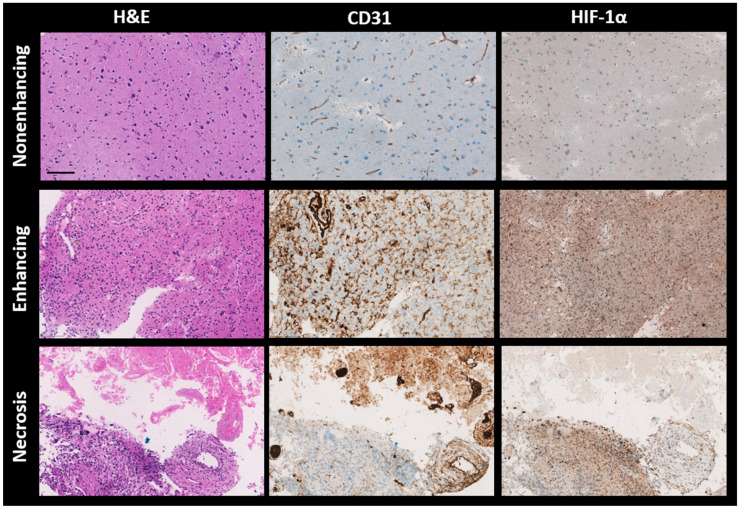
Figure 5.
Representative histological sections of brain tumor tissues from different patients, stained with hematoxylin and eosin (H&E), CD31, and HIF-1α antibodies, are shown from left to right. Top to bottom, the sections are from the nonenhancing, enhancing, and necrotic volumes of interest (VOIs) of brain metastasis (P5), glioblastoma (P10), and glioblastoma (P8), respectively. H&E staining colors nuclei dark blue and cytoplasm pink, while both CD31 and HIF-1α stain nuclei light blue. CD31 specifically highlights endothelial cells in brown, and HIF-1α similarly marks hypoxia-response elements in brown. H&E—hematoxylin and eosin; CD31—cluster of differentiation 31; HIF-1α—hypoxia-inducible factor 1-alpha, Scale bar: 100 μm.

