
Figure 9.
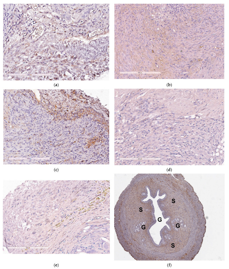
Visual appearance of CD34-stained paraffin sections of endometriotic implants: (a) intact contralateral implant; (b) implant after the treatment with the mock siRNA-complexes; (c) implant after injection of saline; (d) implant after the treatment with the anti-VEGFA siRNA-complexes; (e) implant after the treatment with Dienogest (magnification 200×, bar represents 200 µm); (f) full-size view of intact contralateral EM implant with cyst (G—glandular component; S—stromal component; magnification 40×).