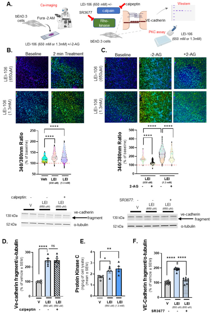
Figure 6.
Increased intracellular calcium level and elevated PKC activity after DAGLα inhibition in bEnD.3 cells. The bEnd.3 cells plated on collagen-coated coverslips were subjected to calcium imaging with the dye Fura-2. After 2-min baseline observation in buffer, cells were treated with LEI-106 (650 µM or 1.3 mM) or vehicle (0.9% DMSO) for 2 min, followed by a subsequent 2-min washout phase. In a separate experiment, cells received buffer spiked with 2-AG (600 pmol/coverslip) during the washout phase. Images used for analysis were captured at the beginning of the treatment application and after removal of the treatment, approximately 2 min apart. In a separate experiment, protein kinase C (PKC) activity was measured by ELISA in bEnD.3 cells after LEI-106 treatment. bEnD.3 cells were treated with calpain inhibitor, calpeptin (10 µM) or Rho-kinase inhibitor, SR3677 (100 nM) 30 min prior to the application of LEI-106 (650 µM), then subjected to Western blot to detect changes in the fragmentation of VE-cadherin caused by DAGLα inhibition. (A) Schema of experimental setting. (B) Representative calcium images of bEnD.3 cells at baseline, then treated with two different doses of LEI-106 (650 µM, 1.3 mM). LEI-106 at both doses significantly increased the intracellular calcium levels compared to vehicle control (LEI-106-650 µM vs. vehicle: p < 0.0001, LEI-106-1.3 mM vs. vehicle: p < 0.0001, as assessed by one-way ANOVA with Tukey post-test, F(3,3295) = 482.1). All values are % of baseline obtained from three independent experiments using three coverslips in each. In each coverslip, 100 cells were analyzed by FIJI Image J software 1.54. **** p < 0.0001 compared to vehicle control. Red dashed line represents the baseline. (C) Representative images showing intracellular calcium levels in bEnD.3 cells treated with LEI-106 (650 µM or 1.3 mM) with or without 2-AG. The application of 2-AG significantly reduced the elevation of intracellular calcium caused by LEI-106 treatment (LEI-106-650 µM vs. LEI-106-650 µM + 2-AG: p < 0.0001, LEI-106-1.3 mM vs. LEI-106-1.3 mM + 2-AG: p < 0.0001, as assessed by one-way ANOVA with Tukey post-test, F(3,2700) = 889.9). All data are shown as % of baseline obtained from three independent experiments using three coverslips in each. In each coverslip, 100 cells were analyzed by Image J software. **** p < 0.0001 compared to vehicle control. Red dashed line represents the baseline. (D) Representative image of Western blot showing VE-cadherin expression in bEnD.3 cells treated with LEI-106 with or without calpain inhibitor, calpeptin (10 µM). α-tubulin was used as a loading control. The calpain inhibitor did not significantly change the fragmentation of VE-cadherin caused by LEI-106 treatment (LEI-106-650 µM vs. LEI-106-650 µM + calpeptin: p > 0.9999, as assessed by one-way ANOVA with Tukey post-test, F(4,20) = 84.40). All data are shown as % of vehicle-treated ± SEM (n = 5–6/condition). **** p < 0.0001, ns = non-significant. (E) The assessment of PKC activity by ELISA showed that LEI-106 treatment (15 min) significantly increased the activity level of PKC compared to vehicle control (LEI-106-650 µM vs. vehicle: * p = 0.0289, t(8) = 2.658; LEI-106-1.3 mM vs. vehicle: ** p = 0.0099, t(8) = 3.362 as assessed by unpaired t-test). Data are presented as the mean of PKC in ng/µg of cell lysate ± SEM (n = 5/group). (F) Representative image of immunoblot targeting VE-cadherin and α-tubulin as loading control in bEnD.3 cells treated with LEI-106 (650 µM) with or without Rho-kinase inhibitor, SR3677 (100 nM). The pretreatment (30 min) with Rho-kinase inhibitor significantly mitigated the fragmentation of VE-cadherin caused by LEI-106 (LEI-106-650 µM vs. LEI-106-650 µM + SR3677: p < 0.0001, as assessed by one-way ANOVA with Tukey post-test, F(2,23) = 110.3). All data are shown as % of vehicle-treated ± SEM (n = 8–10/condition). **** p < 0.0001, compared to the LEI-106 (650 µM) treatment.

