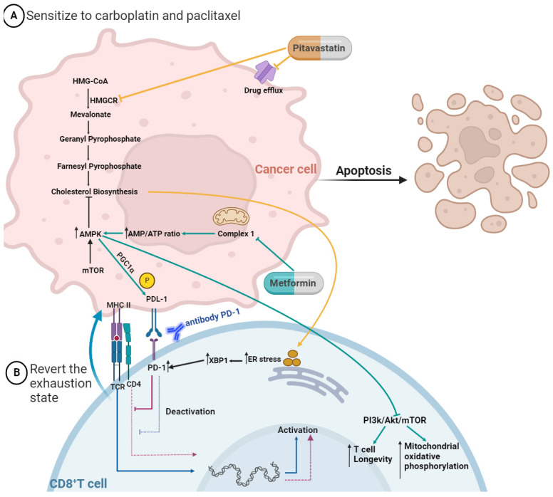
Figure 3.
Mechanisms by which pitavastatin and metformin act to (A) turn cancer cells sensitive to chemotherapy and (B) help to revert the exhaustion state of CD8+ T cells. The pitavastatin blocks drug efflux pumps and inhibits HMGCR, leading to a cascade of inhibition, including cholesterol biosynthesis. The uptake of cholesterol by the exhausted CD8+ T cells increases the stress of the endoplasmic reticulum (ER), leading to the augmented expression of transcription factor X-box binding protein 1 (XBP1), which increases the expression of inhibitory receptors (PD-1). The metformin inhibits the respiratory-chain complex 1, which mediates the activation of adenosine monophosphate-activated protein kinase (AMPK), which inhibits the mammalian target of rapamycin (mTOR) and its downstream signaling pathways. This leads to increased T cell longevity and mitochondrial oxidative phosphorylation. AMPK activation induces PPAR-gamma coactivator 1α (PGC1α), which increases mitochondrial activity and synergistically suppresses tumor growth by phosphorylation of programmed cell death protein ligand-1 (PD-L1). CD8+ T cells are immune active when they have the ligation MHC II with TCR/CD4; on the contrary, when they express the receptor PD-1, they are immune inactive. So, for example, using an antibody to PD-1 allows the inhibition of ligation to PD-1 with PDL-1, and the CD8+ T cell stays active. Figure created in BioRender.com (accessed on 24th of November 2023).

