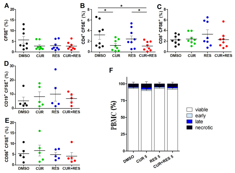
Figure 2.
Effect of low-dose CUR and RES on PBMC proliferation and cell death. (A–E) Cell proliferation of resting PBMCs was assessed by flow cytometry using the dilution of CFSE dye after 96 h of treatment with DMSO, CUR and/or RES (5 µM). The results are presented as the mean ± SD of the frequency of cells subsets in PBMCs from five or eight healthy donors. (A) Total lymphocytes identified based on morphological characteristics on FSC/SSC; (B) CD3+CD19−CD14−CD4+ helper T lymphocytes, (C) CD3+CD19−CD14−CD8+ cytotoxic T lymphocytes, (D) CD3−CD14−CD19+ B cells and (E) CD3−CD19−CD14−CD56+ NK cells, identified by positive staining for the respective markers. Statistical significance of the effects obtained with CUR and RES, alone or in combination, was calculated with two-tailed unpaired Student’s t test (* p ≤ 0.05). (F) Percentages of viable, necrotic, early and late apoptotic cells after 96 h of treatment with DMSO, CUR and/or RES (5 µM) as assessed by the Annexin V/AAD assay and flow cytometry. Results are expressed as the mean ± SD of the independent analysis of PBMCs from eight healthy donors. Statistical significance of the effects obtained with CUR and RES, alone or in combination, was calculated with one-way ANOVA.

