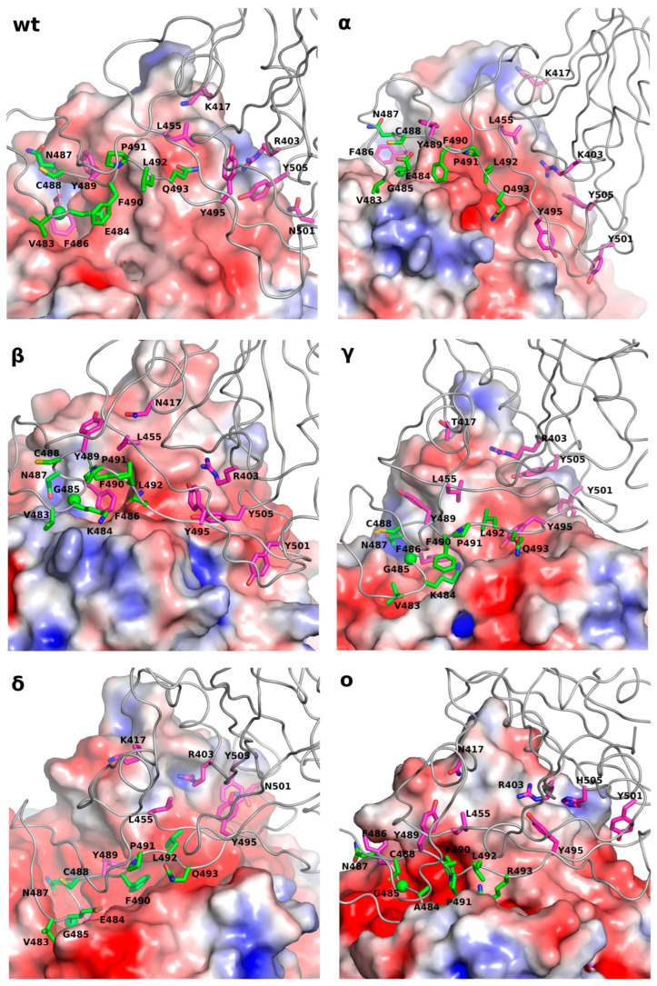
Figure 2.
The intermediate states, identified in all simulated variants of the Receptor-Binding Domain together with Angiotensin-Converting Enzyme-2, were labeled with Greek letters. The most important residues identified as playing a crucial role in the first step of the RBD-ACE2 recognition were labeled in magenta. The residues located in the loop that “sticks” to ACE2 were labeled in green. The Poisson–Boltzmann Surface of the ACE2 protein was generated by using ABPS as implemented in Pymol 2.5.5 software (see methods). The blue region shows the location of positive electrostatic potential, while the red region is the location of negative electrostatic potential. The source of the red spots located in the binding grove comprised the oxygen atoms from the peptide bonds.

