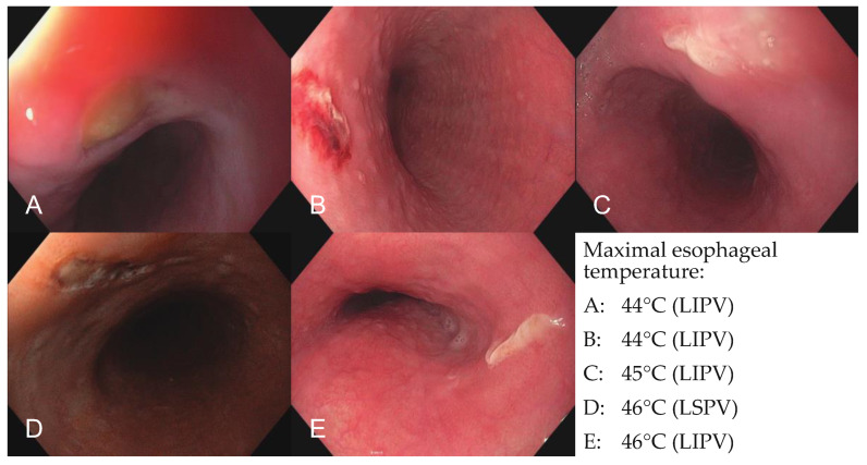
Figure 2.
Esophagoscopic images of endoscopically detected esophageal lesions. Images of all five endoscopically detected esophageal lesions in patients with significant esophageal temperature rise (n = 35) during pulmonary vein isolation with the novel radiofrequency balloon catheter. (A) Type 2a lesion after esophageal temperature rise of 44 °C during ablation of the LIPV, (B) type 1 lesion after esophageal temperature rise of 44 °C during ablation of the LIPV, (C) type 2a lesion after esophageal temperature rise of 45 °C during ablation of the LIPV, (D) type 1 lesion after esophageal temperature rise of 46 °C during ablation of the LSPV, (E) type 2a lesion after esophageal temperature rise of 46 °C during ablation of the LIPV. LSPV = left superior pulmonary vein; LIPV = left inferior pulmonary vein.

