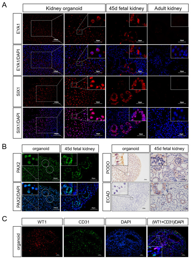
Figure 5.
Gene expression in 3D porcine kidney organoids. (A) Expression of renal progenitor cell markers in the organoids on day 21 and fetal porcine kidney on day 45. Renal progenitor cells markers (EYA1, SIX1) were measured by immunofluorescence test (EYA1 was localized in the cytoplasm, SIX1 was localized in the nucleus). (B) Expression of mature renal cell markers in the organoids on day 21 and fetal porcine kidney on day 45. Mature nephron components markers (PAX2, E-CAD, PODO) were measured by immunofluorescence test and immunohistochemistry test (the circular dotted lines are tubule-like structures; PAX2 was localized in the nucleus; and PODO and ECAD were localized to the cell membrane). (C) Whole-mount co-staining of the organoids for WT1 and CD31 on day 21 (WT1 was localized in the nucleus and CD31 was localized to the cell membrane) (scale bar: 50 μm).

