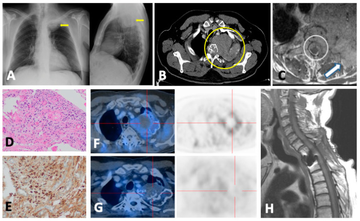
Figure 11.
A 68-year-old male who was admitted to the emergency department for subacute left infraclavicular pain that radiates to the left upper limb. (A) Chest X-ray revealed a mass in the left pulmonary apex (arrows). (B) Chest CT confirmed the presence of a mass in the left pulmonary apex (circle) with mediastinal extension, second left rib destruction, infiltration, and partial destruction of T2 vertebra with probable epidural soft tissue mass and soft tissue component in the adjacent extra thoracic musculature. The differential diagnosis was Pancoast tumor, metastasis of unknown neoplasm, lymphoma, and multiple myeloma/plasmacytoma. (C) Gadolinium-enhanced T1FSWI confirmed the solid nature of the lesion (arrow) and the presence of an epidural soft tissue mass causing cord compression (circle). (D) Histological (hematoxylin–eosin) and (E) immunochemical examinations revealed a clonal proliferation of plasma cells consistent with plasmacytoma. (F) Staging FDG-PET/CT evidenced hypermetabolism of the apical lesion with probable necrotic/hemorrhagic central component without distant skeletal uptakes or additional soft tissue masses. The patient was treated with decompressive surgery, RT, and chemotherapy (T1WI in (H)). (G) One-year follow-up PET/CT showed metabolic response with persistence of tumor.

