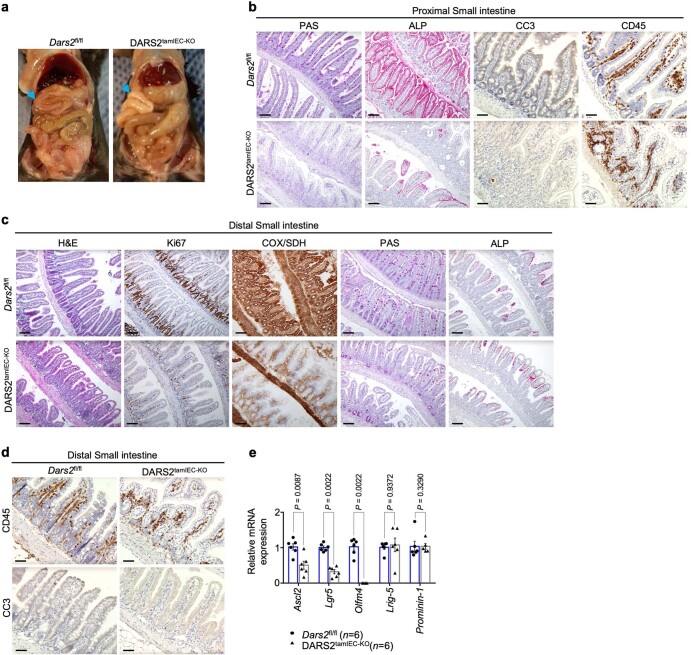
Extended Data Fig. 6. Impaired IEC proliferation, stemness and differentiation upon tamoxifen-inducible DARS2 ablation in IECs of adult mice.
a, Representative pictures from necropsy examination of Dars2fl/fl (n = 4) and DARS2tamIEC-KO (n = 4) mice sacrificed 8 days after the last tamoxifen injection. Blue arrows indicate the proximal SI appearing white in DARS2tamIEC-KO mice. b, c, d Representative microscopic pictures of proximal SI sections stained with PAS and ALP and immunostained with CC3 and CD45 (b) distal SI sections stained with H&E, COX/SDH, PAS, ALP or immunostained with Ki67 (c) and distal SI sections immunostained with CC3 and CD45 from Dars2fl/fl and DARS2tamIEC-KO (d). Scale bar, 50 μm (b, c, d). e, Graph depicting mRNA expression levels of stem cell genes in distal SI of Dars2fl/fl (n = 6) and DARS2tamIEC-KO (n = 6) mice measured with RT-qPCR and normalized to Tbp. In e, dots represent individual mice, bar graphs show mean ± s.e.m. and P values were calculated by two-sided non-parametric Mann-Whitney U test. In b, c and d, histological images shown are representative of the number of mice analysed as indicated in Supplementary Table 4.

