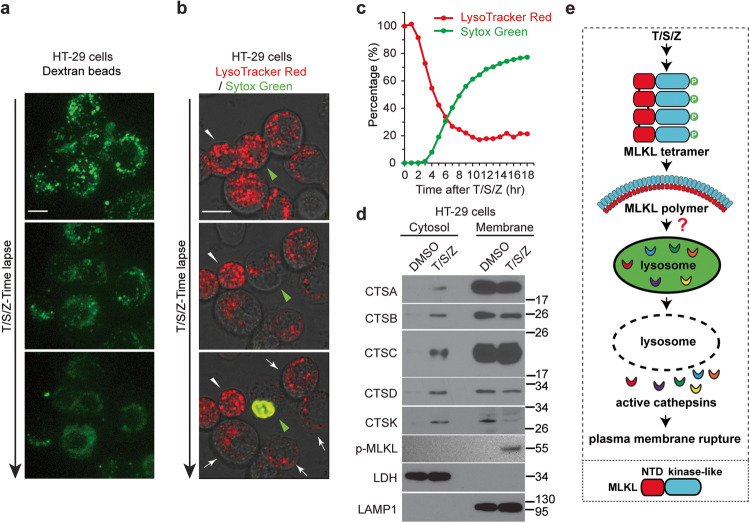
Fig. 1. Lysosomal membrane permeabilization precedes plasma membrane rupture and active cathepsins are released into cytosol during necroptosis.
a HT-29 cells were preloaded with 10 kDa Green Dextran beads overnight. Live cell imaging was recorded after treatment with TNF (T), Smac-mimetic (S) and Z-VAD-FMK (Z). Scale bar, 10 μm. b HT-29 cells were stained with 1 μM LysoTracker Red DND-99 for 2 h followed by 3 washes with PBS. Cells were then treated with 1 μM Sytox Green and T/S/Z, followed by live cell imaging. Scale bar, 10 μm. The green arrowhead identifies a cell undergoing necroptosis and the white arrowhead marks a relatively normal neighboring cell. c HT-29 cells were seeded at a density of 2000 cells/well and stained with LysoTracker Red and Sytox Green. The cells were then treated with DMSO or T/S/Z and florescent images were captured every hour for 18 h. Percentage of red and green signal intensity at each time point was calculated as described in the methods. d HT-29 cells were fractionated into cytosol and membrane fractions after DMSO or T/S/Z treatment for 4 h, followed by Western blotting with the indicated antibodies. The mature active cathepsins were shown. LDH (lactate dehydrogenase) and LAMP1 served as cytosol and membrane markers respectively. Antibody p-MLKL recognizes phospho-S358 of MLKL. e Working model. Upon activation, phosphorylated MLKL forms tetramers and later polymers, somehow leading to LMP and the release of active cathepsins into cytosol, followed by plasma membrane rupture. Diagram on the bottom shows two domains of MLKL, the N-terminal domain (NTD) and the C-terminal kinase-like domain.

