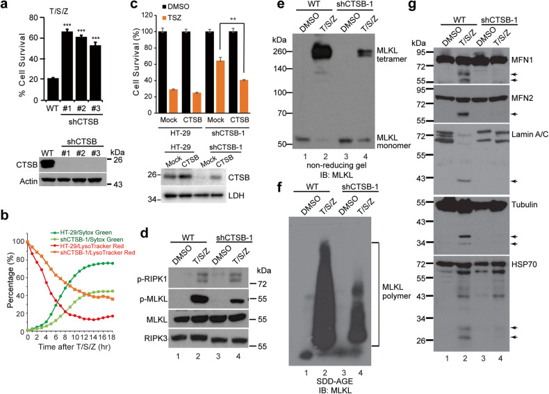
Fig. 5. Loss of CTSB suppresses protein cleavage and necroptosis in HT-29 cells.
a Upper panel, parental HT-29 (WT) and CTSB knockdown (shCTSB) cells were treated with DMSO or T/S/Z for 16 h. Cell survival was measured by CellTiter-Glo assay. ***p < 0.001. Lower panel, Western blotting with antibodies against CTSB and Actin. b LysoTracker Red and Sytox Green staining images for HT-29 or shCTSB-1 cells were recorded and analyzed as in Fig. 1c. c Upper panel, HT-29 or shCTSB-1 cells were transfected with an empty vector or a CTSB expressing plasmid that is resistant to shRNA. Thirty-six hours later, the cells were treated with T/S/Z for 16 h and CellTiter-Glo was performed to assay cell survival. Lower panel, Western blotting with antibodies against CTSB and LDH. d Cells were treated with DMSO or T/S/Z for 4 h and cell lysates were subjected to Western blotting with the indicated antibodies. Cell lysates were analyzed with non-reducing SDS-PAGE (e) or SDD-AGE (f) and probed with an MLKL antibody. g Cell lysates were subjected to Western blotting with the indicated antibodies. Arrows denote cleaved bands.

