. 2023 May 23;42(1):188–201. doi: 10.5534/wjmh.220233

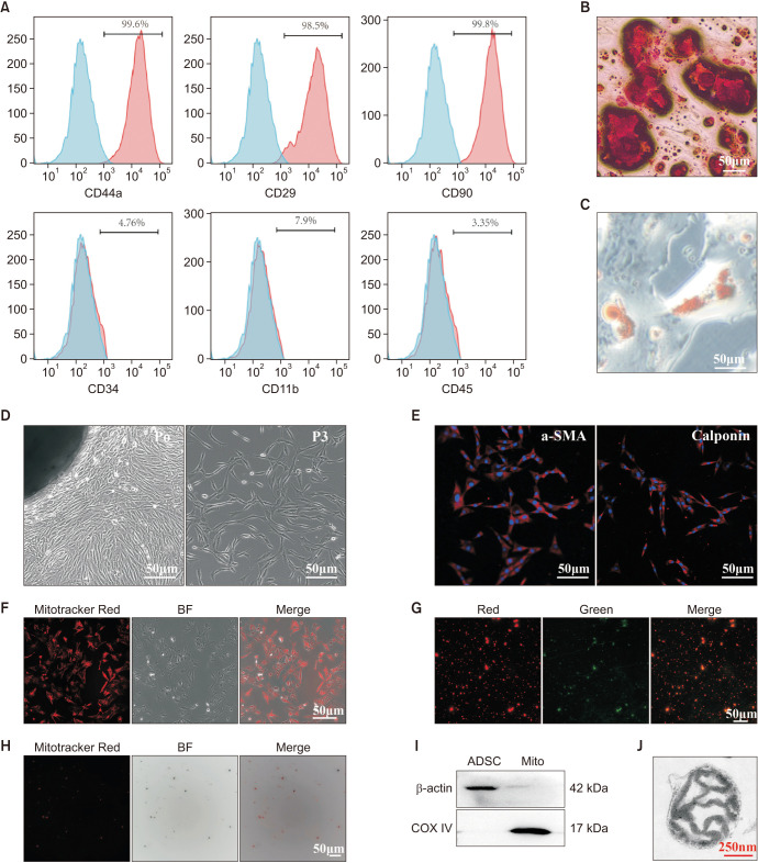
Fig. 1. Characterization of ADSCs, CCSMCs and ADSCs-mito. (A) Flow cytometry showed that ADSCs were positive for mesenchymal stem cells markers (CD44a, CD29, CD90), but not for endothelial or hematopoietic markers (CD11b, CD34, CD45), blue means blank and red means positive percentage for the markers. (B) After induction, ADSCs showed typical osteocyte phenotype (Alizarin Red S staining). (C) After induction, ADSCs showed typical phenotypes of adipocytes (stained with Oil Red O), scale bar, 50 µm. (D) The CCSMCs migrated from penile tissues and grown in a whirlpool-like pattern after passaging, scale bar, 50 µm. (E) Identification of CCSMCs by immunofluorescence with anti-α-SMA and anti-calponin antibody, scale bar, 50 µm. (F) ADSCs were stained with a mitochondrial specific indicator, Mitotracker Red CMXRos. (G) ADSCs-mito was stained with Mitotracker Red CMXRos, scale bar, 50 µm. (H) ADSCs-mito was stained with JC-1, scale bar, 50 µm. (I) The purity of ADSCs-mito was confirmed by COX IV (a mitochondrial marker), ADSCs lysates were used as control. (J) Transmission electron microscopy of isolated ADSCs-mito, scale bar, 250 nm. ADSC: adipose derived mesenchymal stem cell, BF: bright field, CCSMCs: corpus cavernosum smooth muscle cells, ADSCs-mito: ADSCs-derived mitochondria, Mito: mitochondria, α-SMA: alpha smooth muscle actin.

Fig. 1