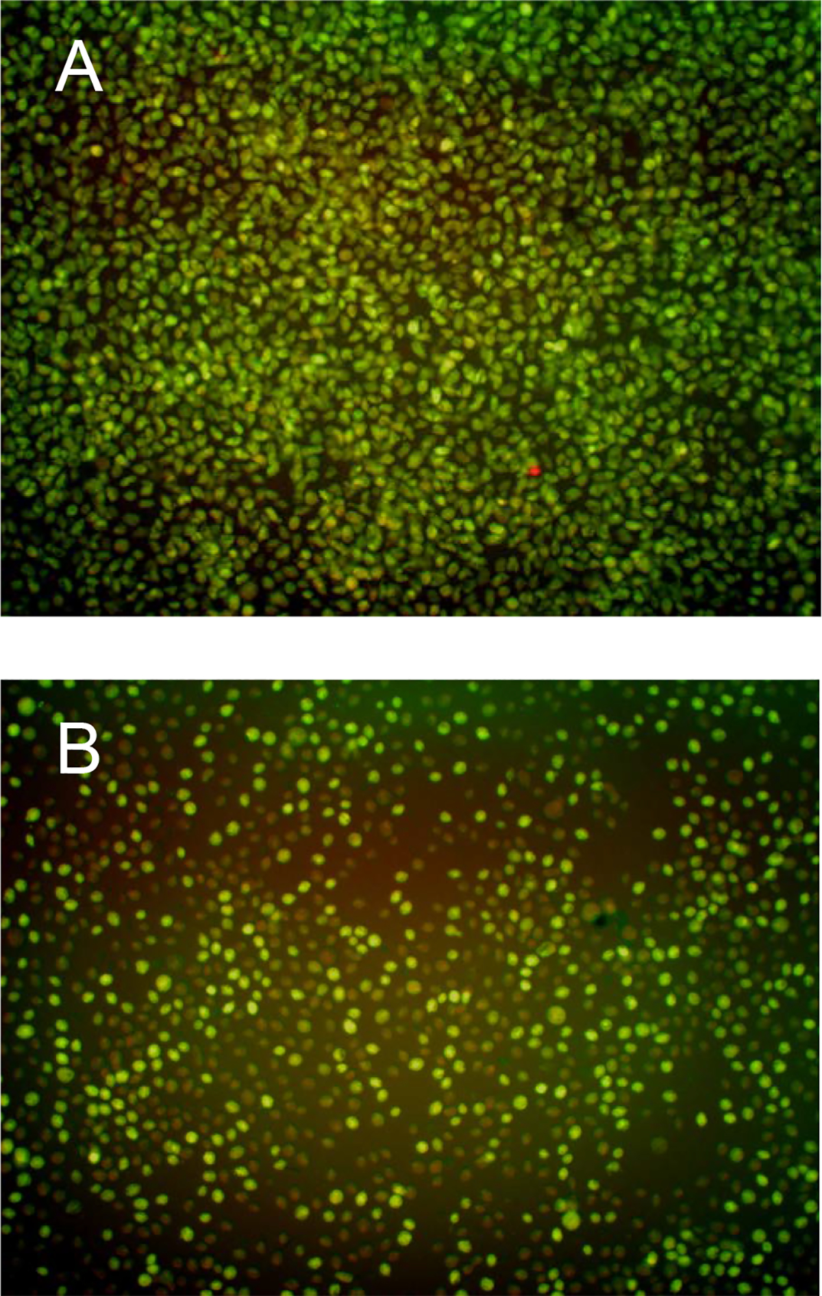
Figure 2.

(A) Sample image produced with the Operetta showing the effects of fixing and staining (0.5% glutaraldehyde, 2.5 μM propidium iodide, and 2.5 μM acridine orange) a confluent T. vaginalis culture at 17 h. (B) Sample image showing the effects of a fungal extract causing partial inhibition of T. vaginalis at 17 h. Note the fewer number of cells, the rounded appearance of the remaining cells, as well as the large number of rust colored cells (the rust color is due to the overlay of green and red emission channels) indicating that a majority of the remaining T. vaginalis cells are dead or dying.
