Fig. 5.
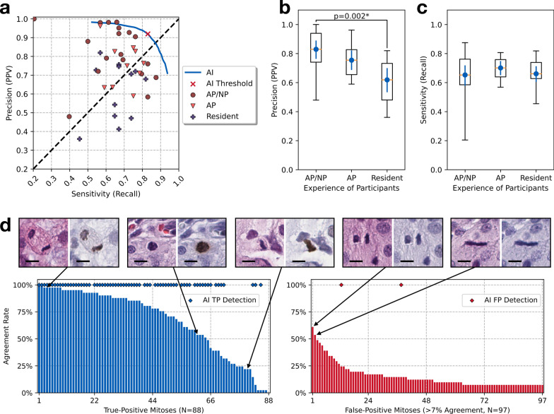
a The precision-recall scatter plot for each participant’s performance in detecting mitoses on the 48 HPF images from the user study. The blue line indicates the precision-recall curve from AI and the marker () indicates AI’s operating point with the threshold cut-off. The diagonal dashed line is the reference where the precision is equal to sensitivity. b The box-whisker plot with the average and 95% confidence interval, showing the precision (PPV) values of the participant groups with different experience levels: AP/NP, AP, and pathology residents. AP/NP participants achieved significantly higher precision than pathology residents (p=0.002, post-hoc Dunn’s test). c Box-whisker plot with 95% confidence intervals, showing the sensitivity (recall) values of the participant groups with different experience levels. No statistical significance was observed. d Bar plots illustrating the agreement rates of participants in identifying ground-truth mitoses and false-positive mitoses, with selected examples (bar=10). The AI detections (both true-positive and false-positive) are shown as the diamond () markers

