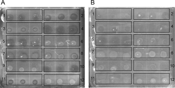
Fig 5.

(A) Pyrene + complex medium and (B) phenanthrene + complex medium top agar assay plate after 5 days of incubation. For each strain, three replicate bacterial spots were plated as follows: (1) R. pomeroyi DSS-3, (2) Citreicella sp. SE45, (3) S. stellata E-37, (4) Bacillus-Clostridium strain SE165, (5) Bacillus-Clostridium strain SE98, (6) M. georgiense DSM 11526, (7) V. natriegens ATCC 14048, (8) Rhodospirillaceae strain EZ35, (9) Alcanivorax sp. strain EZ46, (10) A. macleodii EZ55, (11) Flavobacteriaceae strain EZ40, and (12) E. coli DH5α. Clearing zones appear as dark circles on the media and are visualized after colonies are scraped from the top agar.
