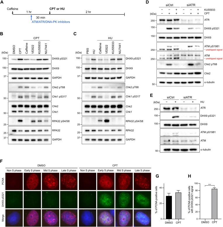
Figure 2.
ATR phosphorylates DHX9 at S321 in response to genotoxic stress. (A–C) A schematic diagram illustrated the experimental sequence designed to investigate the kinase responsible for DHX9 phosphorylation. All indicated inhibitors (caffeine 5 mM, VE822 1 μM, KU55933 10 μM, NU7441 10 μM) were administered for 30 min prior to CPT (1 μM) or HU (2 mM) treatments, except for caffeine, which was administered for 1 h (A). The impact of different inhibitors on the CPT-induced (B) and HU-induced (C) phosphorylation of DHX9 S321 in U2OS cells was assessed by Western blot with indicated antibodies. (D, E) Knockdown of ATR impeded DNA damage-induced S321 phosphorylation. HeLa cells transfected with control and ATR siRNA were treated with CPT (1 μM), CPT plus KU55933 (10 μM) or left untreated for 1 h. Phosphorylation of DHX9, ATM and Chk2 was determined by Western blot (D). The status of S321 phosphorylation in HU-treated cells was determined by Western blot with indicated antibodies (E). (F–H) Immunostaining of PCNA and DHX9 pS321 demonstrated the phosphorylation of DHX9 S321 in S phase. HeLa cells treated with DMSO or CPT (1 μM, 1 h) were subjected to pre-extraction in PBS containing 0.5% Triton X-100, followed by MeOH fixation and immunostaining with indicated antibodies. (F) Representative images of immunostaining. (G, H) Quantification results of PCNA positive cells (G) and PCNA positive cells with DHX9 pS321 signal (H). At least 500 nuclei were quantified and data are presented as the mean ± SD of three independent experiments. Statistical significance was assessed by the Student's t-test (two-sided). *** P < 0.001.

