
Fig. 1.
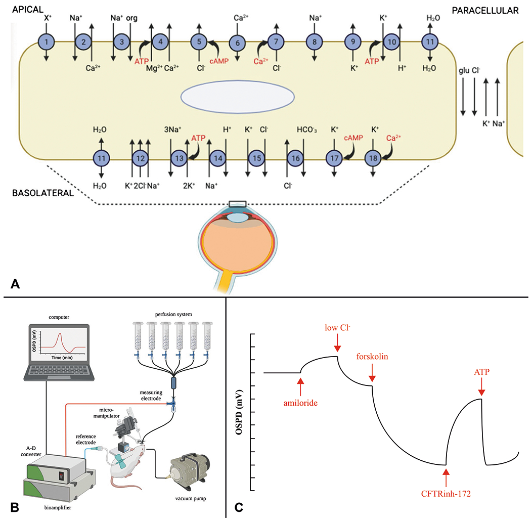
A Schematic diagram of membrane proteins in the ocular surface epithelium (cornea and conjunctiva) involved in the movement of ions via ion channels, pumps, transporters, and receptors [1, 16]. See Table 1 for detailed descriptions. B Schematic of OSPD setup showing the multi-syringe perfusion system to deliver fluid to bathe the ocular surface and the electrical system with measuring electrode in contact with the ocular surface (through the perfusate), subcutaneous reference electrode, and high impedance bioamplifier to measure the mV electrical potential. C Representative OSPD tracing showing PD changes from sequential perfusion exchanges in a typical protocol to study chloride transport