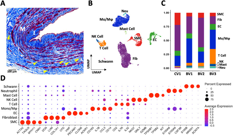
Fig 1. Wall structure and cell composition of upper arm veins.
A) Representative Masson’s trichrome stained cross-section of a basilic vein, indicating the intimal (I), medial (M), and adventitial (A) layers. Cells are stained in red while extracellular matrix appears in blue. Vasa vasorum are indicated by yellow arrowheads. B-C) Uniform manifold approximation and projection (UMAP) plot of 20,006 cells isolated from 4 veins (1 cephalic [CV1], 3 basilic [BV1-3]) and proportion of cell types per vein sample. D) Markers used for cell cluster identification.

