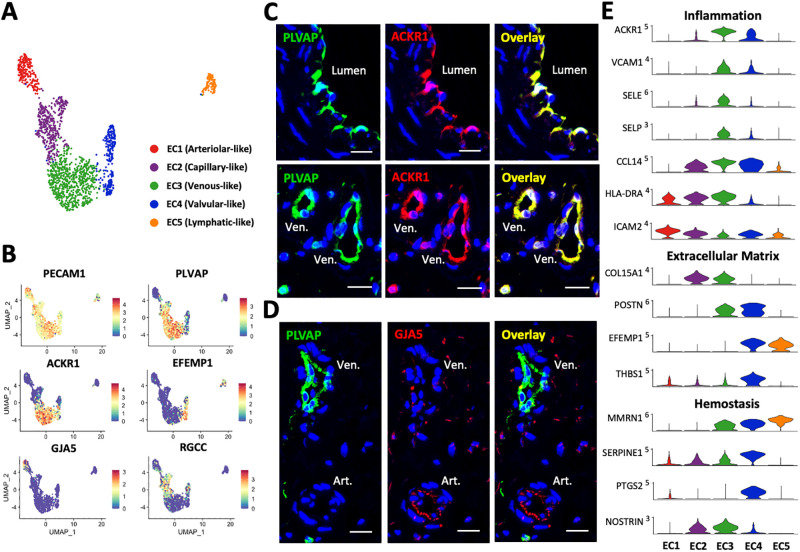
Fig 2. Endothelial cells (EC) in upper arm veins.
A) Focused UMAP of 1,695 ECs isolated from upper arm veins. B) Feature plots indicating normalized expression levels of markers characteristic of the 5 EC populations. C-D) Identification of venous (PLVAP+ACKR1+) and arteriolar (PLVAP-GJA5+) ECs in the main lumen of veins and vasa vasorum by immunofluorescence. Ven. = venule; Art. = arteriole. Scale bars represent 20 μm in all panels. E) Violin plot representation of expression differences among the 5 EC populations.

