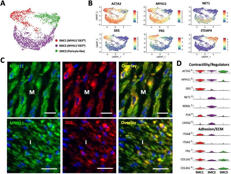
Fig 3. Smooth muscle cells (SMC) and pericytes in upper arm veins.
A) Focused UMAP of 2,298 SMCs isolated from upper arm veins. B) Feature plots indicating normalized expression levels of markers characteristic of the 3 SMC populations. C) Localization of SMC1 (MYH11+DEShi) and SMC2 (MYH11+DESlo) cells in the intima (I) and media (M) by immunofluorescence. Scale bars represent 20 μm in all panels. D) Violin plot representation of expression differences among the 3 SMC populations.

