. 2023 Dec 19;12:e85096. doi: 10.7554/eLife.85096

Figure 3. Predicted alkaloid-binding globulin (ABG) structure and binding pocket resembles that of other small molecule binding serpins.

(A) AlphaFold structure predicted with the protein sequence of the Oophaga sylvatica ABG, with color representing model confidence and predicted binding pocket based on molecular docking simulation indicated with a black box. (B) Crystal structure for rat SerpinA6/corticosteroid-binding globulin (CBG), with the cortisol molecule shown in orange (PDB# 2V95). (C) Crystal structure for tree frog Boana punctata biliverdin-binding serpin (BBS), with biliverdin shown in orange (PDB# 7RBW). (D) Close-up of predicted binding pocket of pumiliotoxin (PTX) in O. sylvatica ABG, with residues proximal to PTX highlighted in magenta. The structure of PTX is indicated on the top right. (E) Close-up of cortisol binding in CBG (PDB# 2V95), with proximal residues highlighted in magenta. Cortisol structure is displayed on the top right. (F) Close-up of biliverdin binding in BBS (PDB# 7RBW), with some proximal residues highlighted in magenta. Biliverdin structure is shown on the top right.

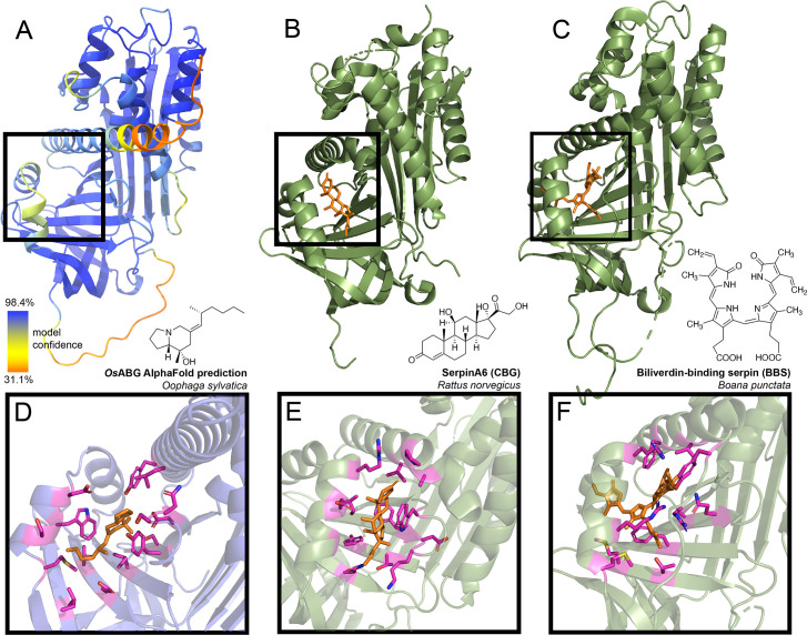
Figure 3—source data 1. Raw data for the structure prediction shown in Figure 3.

Figure 3.

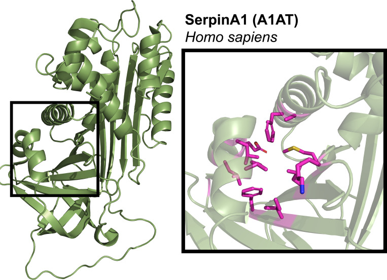
Figure 3—figure supplement 1. Structure of A1AT protein.

Figure 3—figure supplement 1.

Crystal structure for human SerpinA1/alpha-1-antitrypsin (PDB# 1HP7) contains the structural elements of other serpin-binding pockets (black box), however is not documented to have small molecule binding capabilities. Black box shows close-up of A1AT (PDB# 1HP7), with pocket residues highlighted in magenta.
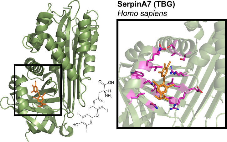
Figure 3—figure supplement 2. Structure and binding pocket of thyroxine-binding globulin (TBG) protein.

Figure 3—figure supplement 2.

Crystal structure for human SerpinA7/TBG (PDB# 2RIW) binds thyroxine (orange) in the same structural pocket as other serpins. Black box shows close-up of thyroxine-binding pocket of TBG (PDB# 2RIW), with proximal residues highlighted in magenta. Thyroxine structure is displayed on the top right.
Figure 3—figure supplement 3. Alignment of binding pocket residues.

Figure 3—figure supplement 3.

Alignment of proximal residues (within 5 angstroms of small molecule) across small molecule binding serpins OsABG, biliverdin-binding serpin (BBS, PDB# 7RBW), corticosteroid-binding globulin (CBG, PDB# 2V95), and thyroxine-binding globulin (TBG, PDB# 2RIW) shows that most residues that may be involved in coordinating small molecule binding are not conserved, despite the structural conservation of the binding pocket. Percentages indicate the total percent identity of the protein sequences, the small number above each residue indicates the position of that amino acid in the protein sequence. Only proximal residues are shown, blank spaces are not representative of any specific sequence or spacing.
Figure 3—figure supplement 3—source data 1. Raw data for the alignment shown in Figure 3—figure supplement 3.