. 2023 Dec 19;12:e85096. doi: 10.7554/eLife.85096

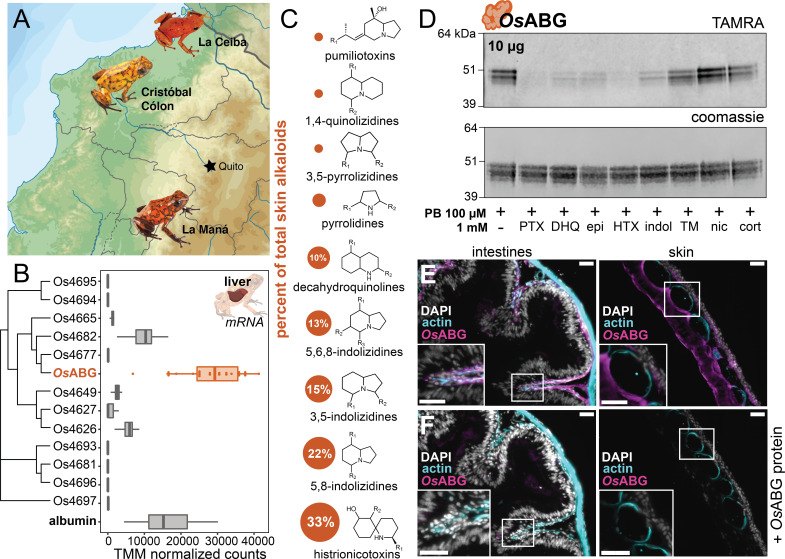
Figure 6. OsABG is expressed in the liver and binds ecologically relevant alkaloids.

(A) Wild Oophaga sylvatica were collected across three locations in Ecuador, n = 10 per location. (B) The liver expression level of OsABG was higher than that of other members of the serpinA family found in the genome, and of albumin. (C) Dorsal skin alkaloids fell into nine different classes, with the size of the circle representing the averaged percent of total skin alkaloid load. (D) Photoprobe binding with recombinantly expressed OsABG was competed by pumiliotoxin (PTX), decahydroquinoline (DHQ), epi, a histrionicotoxin-like compound (HTX), and indolizidine ring without R groups (indol), and slightly by a mixture of skin toxins from the wild specimens (TM). Photoprobe binding was not competed by nicotine (nic) or cortisol (cort). (E) Custom anti-OsABG antibody staining (magenta) in the skin and intestines, with actin stain (blue) and 4',6-diamidino-2-phenylindole (DAPI, shown in white). (F) Pre-incubation of anti-OsABG with purified OsABG protein in the skin and intestines shows loss of OsABG staining, indicating specific staining activity. White bars represent 50 μm.

Figure 6—source data 1. Raw data for the gene expression, gels, and immunohistochemistry shown in Figure 6.

Figure 6.

Figure 6—figure supplement 1. OsABG expression in intestines, liver, and skin.
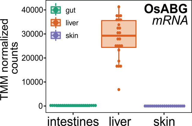
Figure 6—figure supplement 1.

Comparison of mRNA expression levels across tissues found high expression in the liver, and low to no expression in the skin and intestines.
Figure 6—figure supplement 1—source data 1. Raw data for the gene expression data shown in Figure 6—figure supplement 1.
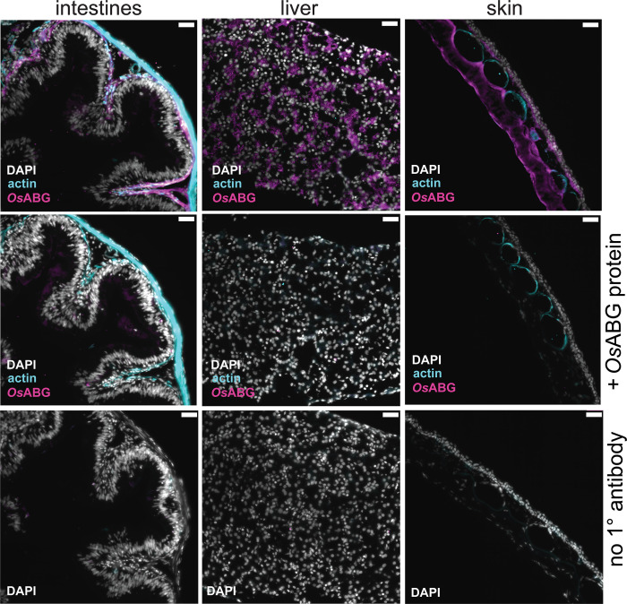
Figure 6—figure supplement 2. OsABG protein presence in intestines, liver, and skin.

Figure 6—figure supplement 2.

Immunohistochemical staining of O. sylvatica intestines, liver, and skin with custom anti-OsABG primary antibody, anti-actin, and DAPI. Second row shows staining of O. sylvatica intestines, liver, and skin with custom anti-OsABG primary antibody pre-incubated with OsABG protein, anti-actin primary antibody, and DAPI stain. Third row shows negative control staining of O. sylvatica intestines, liver, and skin with no primary antibodies added.
Figure 6—figure supplement 2—source data 1. Raw data for the immunohistochemistry shown in Figure 6—figure supplement 2.
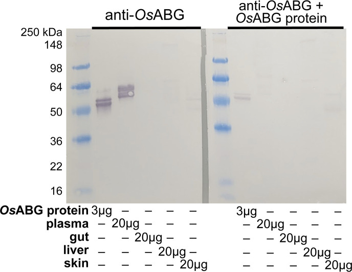
Figure 6—figure supplement 3. Western blot of anti-OsABG protein.

Figure 6—figure supplement 3.

Western blot using custom anti-OsABG antibody against purified OsABG protein, O. sylvatica plasma, intestines, liver, and skin. The second blot has the same conditions but the primary antibody was pre-incubated with purified OsABG protein prior to blotting, to test the specificity of the antibody.
Figure 6—figure supplement 3—source data 1. Raw data for the western blot shown in Figure 6—figure supplement 3.