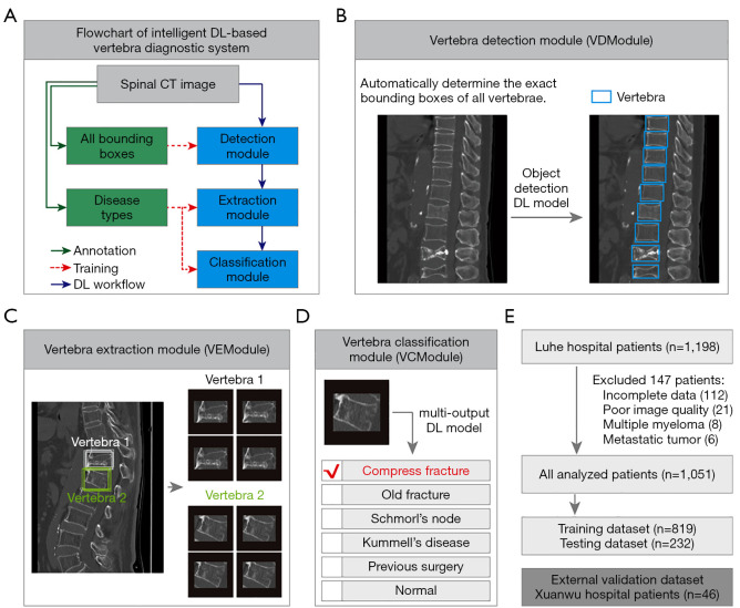
Figure 1.
Architecture of intelligent DL-based vertebra diagnostic system. (A) Flowchart for the development of a DL-based vertebra diagnostic system. Expert annotation included two types: labeling all vertebrae by Bboxes for training vertebra detection models and labeling vertebrae with diseases for training vertebra classification models. The DL workflow consists of three modules: a VDModule, a VEModule, and a VCModule. (B) The function of the VDModule. The VDModule processes the clinical CT image and automatically determines the exact Bbox of all vertebrae. Left, clinical CT image. Right, detected vertebrae. The VDModule is developed based on object-detection DL models. (C) The function of VEModule. The VEModule obtains multiple samples from one Bbox of the target vertebra by implementing scaling and random translation, thereby realizing data augmentation. The eight cropped vertebra image patches shown as gray and green Bboxes correspond to vertebrae 1 and 2, respectively. (D) The function of the VCModule. The VCModule is developed based on a multi-output DL model and implements the diagnosis of the input vertebra patch. The vertebra types recognized by VCModule include OVCF, OFs, SN, KD, PS, and normal. (E) Two patient cohorts were used in this study. The training and testing datasets were from Luhe Hospital and the alternative hospital validation dataset was from Xuanwu Hospital. DL, deep learning; CT, computed tomography; VDModule, vertebra detection module; VEModule, vertebra extraction module; VCModule, vertebra classification module; Bboxes, bounding boxes; OVCF, osteoporotic vertebral compression fracture; OF, old fracture; SN, Schmorl’s node; KD, Kummell’s disease; PS, previous surgery.

