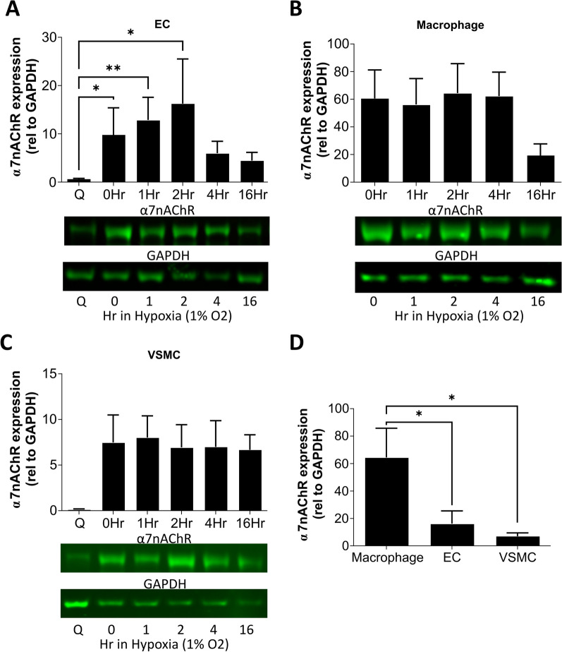
Fig. 1.
Analysis of α7nAChR protein in human cells associated with vascular regeneration and repair showing an endothelial-specific hypoxia response, and the highest α7nAChR expression relative to GAPDH within macrophages. A Western blot quantification of human endothelial cell (HUVEC) α7nAChR expression during quiescent conditions (Q), normoxic proliferating conditions (0Hr) and exposure to 1% hypoxia (1–16 h) in proliferating conditions. N = 6, * = P ≤ 0.05, ** = P ≤ 0.01 using a Kruskal–Wallis test with post hoc Dunn’s. B Western blot quantification of human macrophage (PMA-differentiated THP-1) α7nAChR expression during normoxic conditions and 1% hypoxia. These cells no longer proliferate following PMA differentiation, so no quiescent conditions were used, N = 4–5. C Western blot quantification of vascular smooth muscle cell (human coronary artery) α7nAChR expression during quiescent conditions (Q), normoxic proliferating conditions (0Hr) and exposure to 1% hypoxia in proliferating conditions, N = 4–5. D Direct comparison of α7nAChR expression across the cell types at 2 h in 1% hypoxic conditions, N = 5–6 and * = P ≤ 0.05 using ordinary one-way ANOVA with post hoc Holm-Šidák test. All results are shown as the mean ± SEM and example blots are shown

