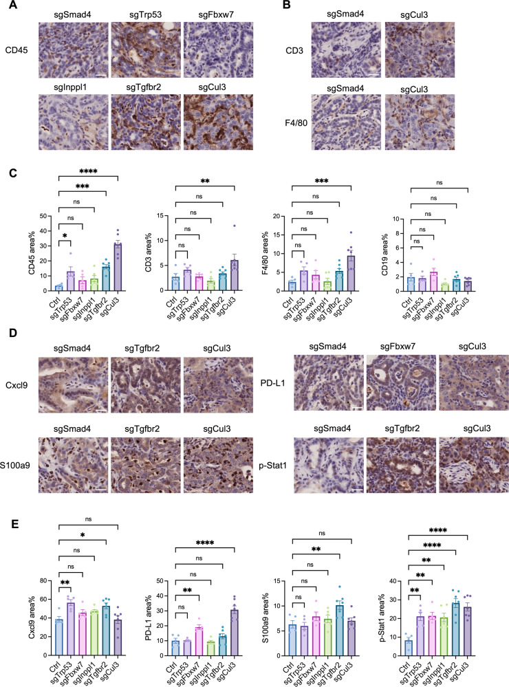
Fig. 3. The tumor microenvironment varies in ICC with different gene mutations.
A, B IHC staining of immune cells in ICC tumors developed in SPC mice. CD45 indicates total immune cells, CD3 indicates T cells, and F4/80 indicates macrophages. C Statistical analysis of immune markers in tumors, each dot indicates one mouse. sgSmad4 was included as a negative control. D, E IHC staining and statistical analysis of immune-related molecules in ICC tumors developed in SPC mice. ns: not significant, *p < 0.05, **p < 0.01, ***p < 0.001, ****p < 0.0001. Scale bar = 25 μm.

