Fig. 4.
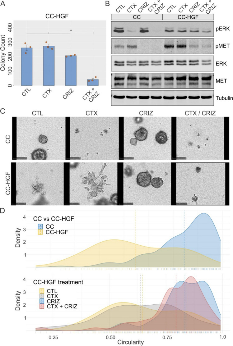
Crizotinib overcomes HGF-induced cetuximab resistance and loss of polarity. A Two thousand CC-HGF cells were seeded in type I collagen and incubated with cetuximab (CTX, 3 μg/ml) or crizotinib (CRIZ, 0.25 μM) for 14 days. Colony counts are plotted as mean ± SEM; * indicates statistically significant differences (one-way ANOVA with Tukey's HSD post hoc test, p < 0.05). B One hundred thousand CC and CC-HGF cells were cultured in type I collagen for seven days and incubated with CTX (3 μg/ml) and/or crizotinib (CRIZ, 0.25 μM) for an additional 48 h then lysed and resolved on SDS-PAGE followed by immunoblotting for proteins as indicated. C Live-cell imaging of CC vs CC-HGF treated with CTX and/or CRIZ: CC and CC-HGF cells imaged for 12 days with treatments indicated. Final image at the end of analysis is displayed. Supplementary movies contain the corresponding full-length movies of imaging of individual colonies under treatment. D Quantification of cystic and spiky morphology of cells grown in 3D for eight days with treatments indicated. Results are quantified as circularity index (see Methods); higher circularity index indicates rounder colonies and lower circularity index indicates spiky colonies. Top panel compares CC and CC-HGF circularity at baseline, and lower panel depicts CC-HGF cells under indicated treatment conditions. Dotted lines indicate median circularity index for indicated cells and treatment conditions

