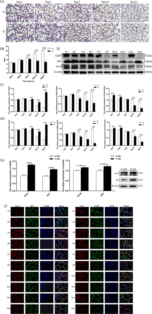
Fig. 1. In BPD, alveolation is impaired, and Sox9 expression level is significantly changed.
A Representative images of H&E staining of lung tissue. Morphology was examined using light microscopy (×200). Scale bar: 50 μm. B Comparison of RAC values in lung tissues. C Relative levels of SPC, AQP5, Sox9 mRNAs in rat lung tissue (n = 6). D Relative levels of SPC, AQP5, Sox9 protein in rat lung tissue (n = 6). E Target band intensity was normalized to β-actin. F Paraffin-embedded tissue sections were double stained with Sox9 (red) + AQP5 (green) immunofluorescence (×400). G Relative levels of SPC, AQP5, Sox9 mRNAs, and protein in the in vitro cell model. Target band intensity was normalized to β-actin. (**P < 0.01, ***P < 0.005, and ****P < 0.001 compared with the control group).

