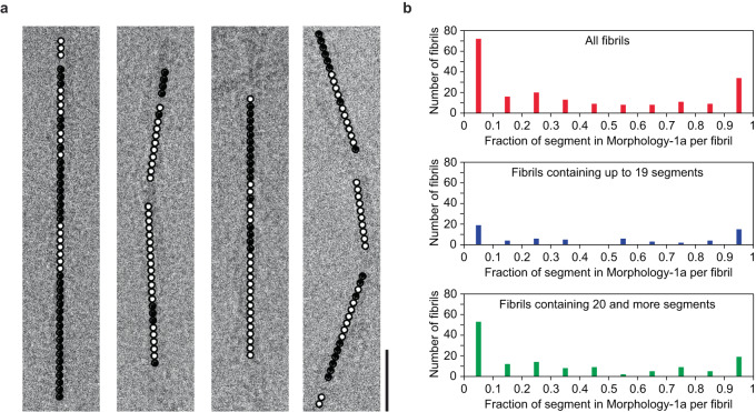
Fig. 3. Evidence for structural breaks in Morphology-1.
a Cropped cryo-EM images show the Morhphology-1a (black dot) and Morphology-1b (white dot) segments which occur clustered into distinct z-axial regions. Morhphology-1a and 1b segments were found to coexist in 132 out of 200 analyzed fibrils. Scale bar: 25 nm. b The fraction of Morphology-1a segments, per fibril (200 fibrils, top). Evaluation of a subset of fibrils with 1–19 segments (64 fibrils, middle) and of fibrils with 20 or more segments (136 fibrils, bottom).

