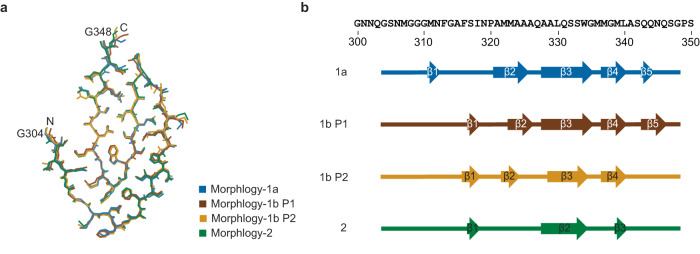
Fig. 4. Fibril protein fold in the three fibril morphologies.
a Super-imposition of the fibril proteins of Morphology-1a, Morphology-1b and Morphology-2 (stacks P1 and P2). The first and the last amino acid residues of the fibril core are labeled. Note that differences in the side chain geometries may have arisen from the modeling. b Sequence of residues Gly300 to Ser350 of TDP-43 drawn side-by-side with schematic representations of the fibril protein core in the different fibril morphologies as indicated in the panel. Arrows indicate β-strands. The color coding of the fibril morphologies is kept consistent in both panels.

