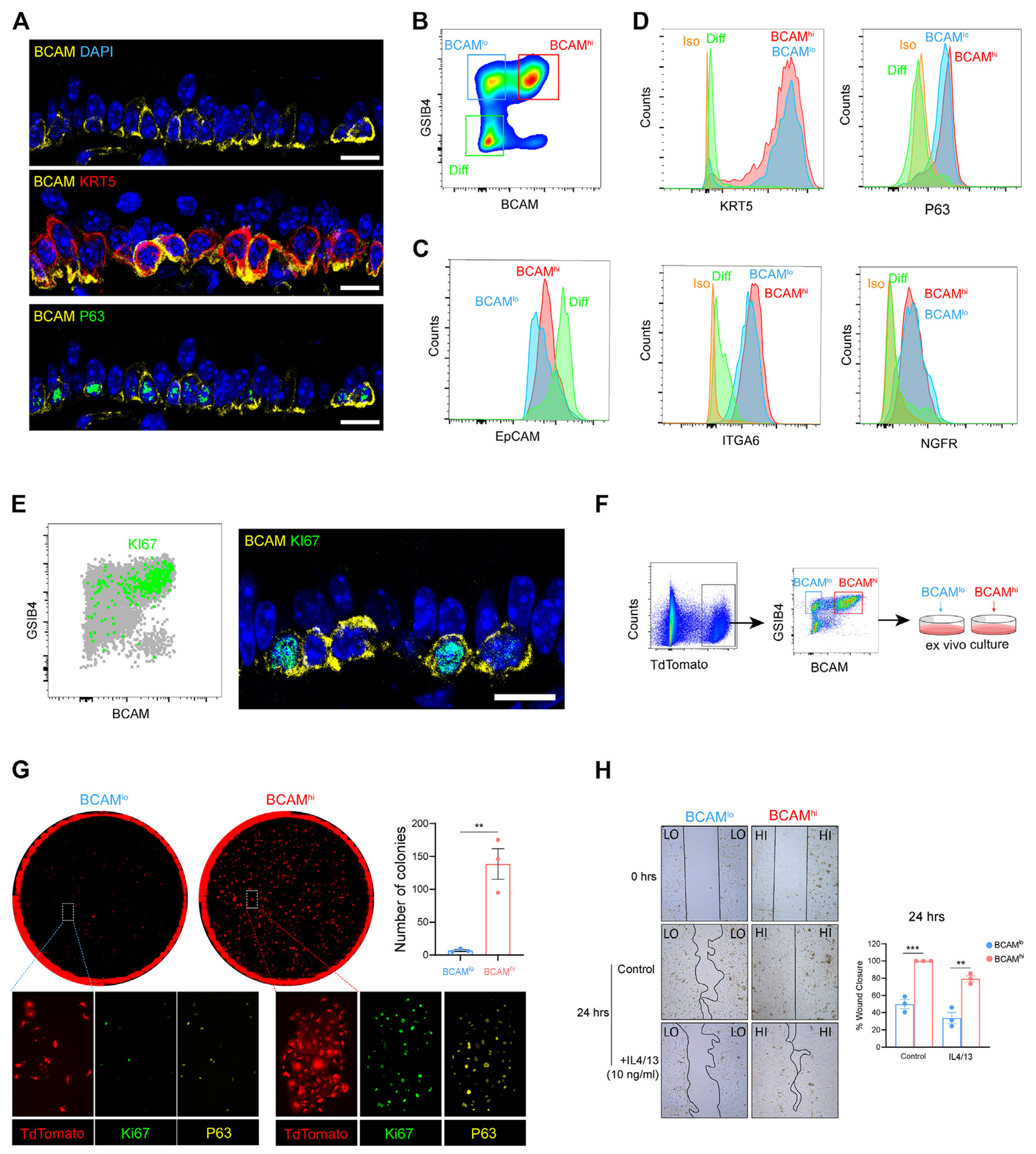
FIG 2.

BCAM marks an airway basal stem cell among KRT5+NGFR+ITGA6+P63+ BCs in the naive murine trachea. A, Representative immunostaining in naive murine trachea. The scale bar represents 10 μm. B, Flow-cytometric panel of BCAMhi BCs, BCAMlo BCs, and differentiated EpCs (Diff). C, Expression of EpCAM in each group. D, Expression of canonical BC markers in each group. E, KI67 expression in naive tracheal airway epithelium assessed by flow-cytometric staining (left) and confocal microscopy (right). The scale bar represents 10 μm. F, Schema depicting the isolation and ex vivo culture of BCAMhi BCs and BCAMlo BCs from KRT5CrerERT2:R26tdTomato mice (for detailed information, see this article’s Methods section in the Online Repository at www.jacionline.org). G, Colony-forming assay on sorted BCAMhi BCs and BCAMlo BCs with immunostaining. Number of colonies was calculated using image J. Data are shown as mean ± SEM (n = 3; **P < .01; unpaired 2-tailed t test). H, Wound-healing assay on sorted BCAMhi BCs and BCAMlo BCs in the presence or absence of 10 ng/mL IL-4 and IL-13. Images were taken at 0 and 24 hours. The percentage of wound closure was calculated using image J. Data are shown as mean ± SEM (n = 3; P < .02; linear regression). All immunostaining is representative of n = 3. DAPI, 4’-6-Diamidino-2-phenylindole.
