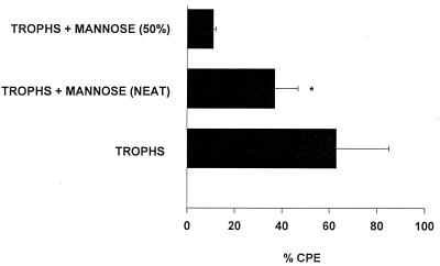
FIG. 4.
Cytolytic activity of supernatants from A. castellanii trophozoites stimulated with mannose. Trophozoites were incubated with 100 mM mannose for 48 h at 35°C. Supernatants were collected, centrifuged, filter sterilized, dialyzed, and added to CHCE cell monolayers. CPE was assessed spectrophotometrically after 48 h. Positive controls consisted of CHCE cell monolayers incubated with 2.5 × 10⁶ trophozoites/ml (trophs). Negative controls consisted of monolayers incubated in medium alone and, by definition, had no CPE (data not shown). ∗, the difference between the undiluted mannose supernatant group (neat) and the medium control (0% CPE; not shown) was significant (P = 0.0078). Trophs + mannose (50%), mannose supernatant diluted 1:2 in complete MEM. Results are expressed as mean ± standard deviation.

