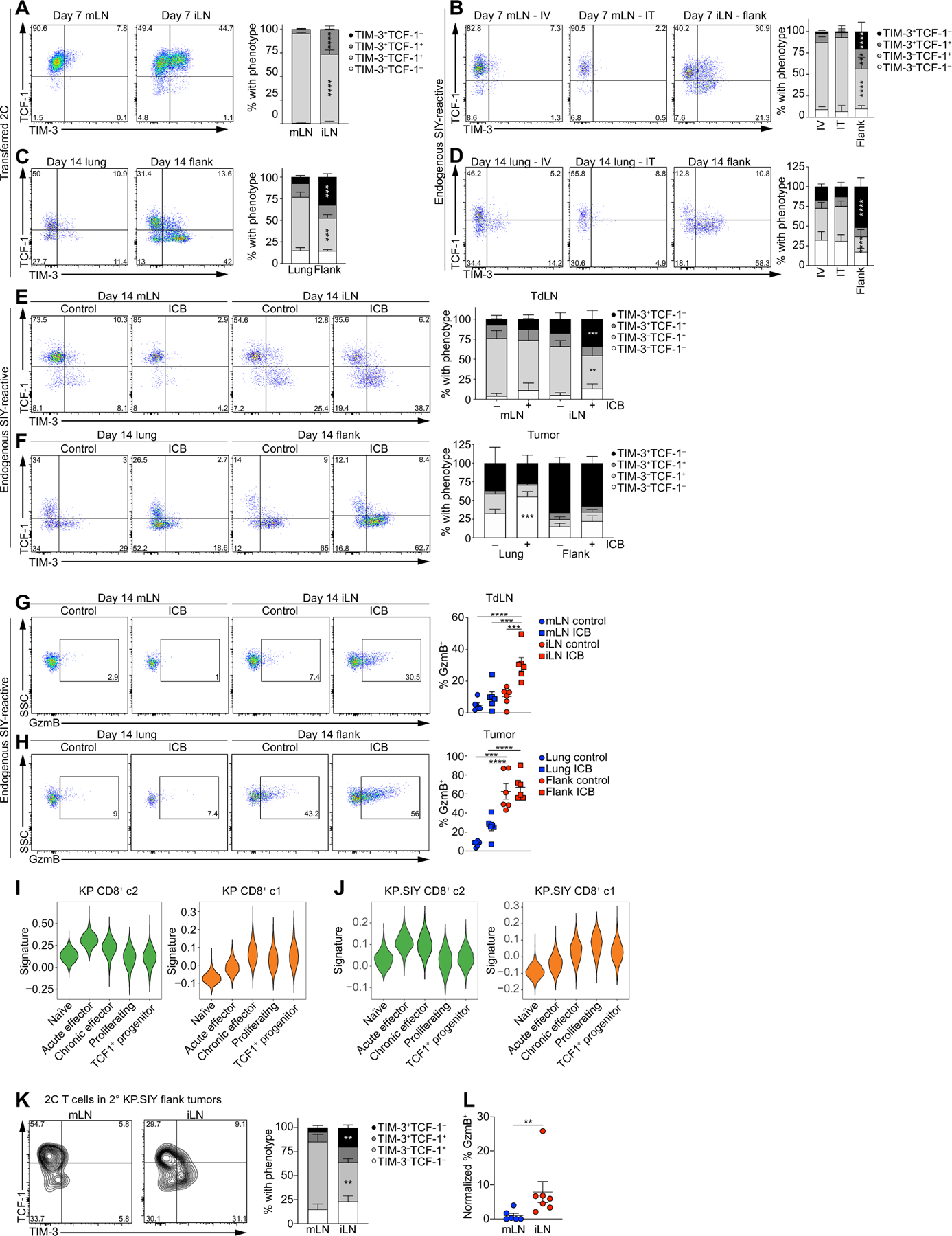
Fig. 5. TLdys T cell state is functionally distinct from conventional Tex T cell state.

(A to D) Flow cytometry analysis of TCF-1 and TIM-3 expression on adoptively transferred 2C T cells in the mLN and iLN of KP.SIY tumor–bearing mice 72 hours after adoptive transfer (10 days after tumor inoculation; n = 14 for mLN and n = 10 for iLN, data pooled from two experiments). (B) Endogenous, SIY-reactive CD8+ T cells in KP.SIY tumor–bearing mice 7 days after intravenous, intratracheal, or subcutaneous inoculation (n = 9 for IV mLN, n = 10 for IT mLN, and n = 9 for iLN, data pooled from three experiments). (C) Adoptively transferred 2C T cells in lung or flank KP.SIY tumors 14 days after tumor inoculation and 7 days after adoptive transfer (n = 6 for lung and n = 5 for flank, data pooled from two experiments). (D) Endogenous, SIY-reactive CD8+ TIL in KP.SIY tumor–bearing mice 14 days after tumor inoculation (n = 6 for all conditions, data pooled from two experiments). (E to H) Analysis of SIY-reactive CD8+ T cells in KP.SIY tumor untreated or treated with ICB given on days 7 and 10 after tumor inoculation. TCF-1 and TIM-3 expression in mLN and iLN (E) and lung and flank tumors (F) 14 days after tumor inoculation (n = 6, data pooled from two experiments). GzmB expression on SIY-reactive CD8+ T cells in (G) mLN and iLN and lung and flank tumors (H) 14 days after tumor inoculation (n = 6, data pooled from two experiments). (I and J) Comparison of CD8+ c2 (green) or CD8+ c1 (orange) signatures from KP parental (I) and KP. SIY (J) datasets with previously published CD8+ T cell signatures from acute and chronic LCMV infection. (K and L) Flow cytometric analysis of TCF1–1, TIM-3 (K), and GzmB (L) expression in serially transferred 2C T cells on day 5 after transfer to RAG2−/− (n = 6 for mLN and n = 7 for iLN, data pooled from two experiments) Data are shown as means ± SEM, and statistical analysis was conducted using an MWU test with **P < 0.01, ***P < 0.001, and ****P < 0.0001 (A to H and K to L).
