Fig. 3.
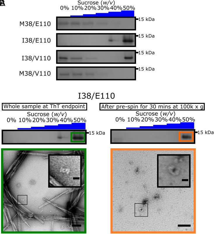
γSyn I38 forms distinct oligomeric species. (A) SDS-PAGE analysis of the γSyn variants at pH 7.5 after zonal density gradient ultracentrifugation at 113,000g for 4 h in a discontinuous sucrose gradient. The entire gels are shown in SI Appendix, Fig. S7 A–D. (B) (Top) SDS-PAGE showing that the γSyn I38/E110 species which do not pellet at 100,000g after 30 min in buffer are found exclusively in the 50% (w/v) sucrose fraction following zonal density gradient ultracentrifugation. The entire gel is shown in SI Appendix, Fig. S7E. (Bottom) Negative stain TEM micrographs representing the 50% (w/v) sucrose fraction of γSyn I38/E110 before (Left, Scale bar, 100 nm) and after removal of fibrillar material (Right, Scale bar, 50 nm). A zoom of each sample is shown Inset. (Scale bars, 20 nm.)

