Figure 2.
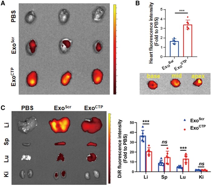
In vivo biodistribution of ExoCTP. MI mice were intravenously injected with DiR-labeled ExoCTP for in vivo tracking. Different organs were dissected 6 h post-injection (A) ex vivo optical imaging and semi-quantification for infarcted hearts (n = 6). (B) Ex vivo imaging of heart sections from base to apex in ExoCTP group. (C) Ex vivo optical imaging and semi-quantification of other major organs (n = 6). All data are represented as mean ± SD. Two-tailed unpaired Student’s t test was utilized. ns: not significant, ***P < 0.001.

