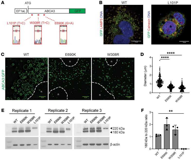
Figure 4. Protein trafficking and lamellar body phenotypes are recapitulated in response to lentiviral forced overexpression of mutant and normal ABCA3:GFP fusion proteins in A549 cell lines.
(A) Schematic showing locations and sequencing confirmation of 3 introduced ABCA3 mutations in 3 separate lentiviral vectors (each vector contains no or only 1 mutation, denoted by *) used to transduce A549 cells. Each of the 4 lentiviral vectors expressing WT or mutant ABCA3:GFP fusion constructs were driven by a constitutively active promoter EF1aL. *, Site directed mutagenesis of missense mutations confirmed by sequencing. (B) Representative confocal fluorescence microscopy of A549 cells expressing WT and L101P ABCA3:GFP fusion proteins (green) costained with lamellar body marker LAMP3 (red, left) and ER marker calnexin (red, right). Nuclei (blue). Scale bars: 10 μm. (C) Representative live-cell high resolution confocal images showing smaller GFP+ intracellular vesicles formed in cells expressing E690K or W308R mutant ABCA3:GFP fusion proteins compared with WT fusion protein. Nucleus (n), dotted lines. Scale bars: 5 μm. (D) Quantitation of the diameter of GFP+ intracellular vesicles in indicated A549 samples. ****P ≤ 0.0001 by 1-way ANOVA with Tukey’s multiple comparisons test. (E) Western blot using antibody against GFP to compare 220 kDa to 180 kDa protein cleavage of WT versus E690K, W308R, and L101P mutant ABCA3:GFP fusion proteins over 3 replicates. (F) Quantification of percentage of total ABCA3:GFP protein cleaved in each indicated sample. **P ≤ 0.01 by 1-way ANOVA with Tukey’s multiple comparisons test.

