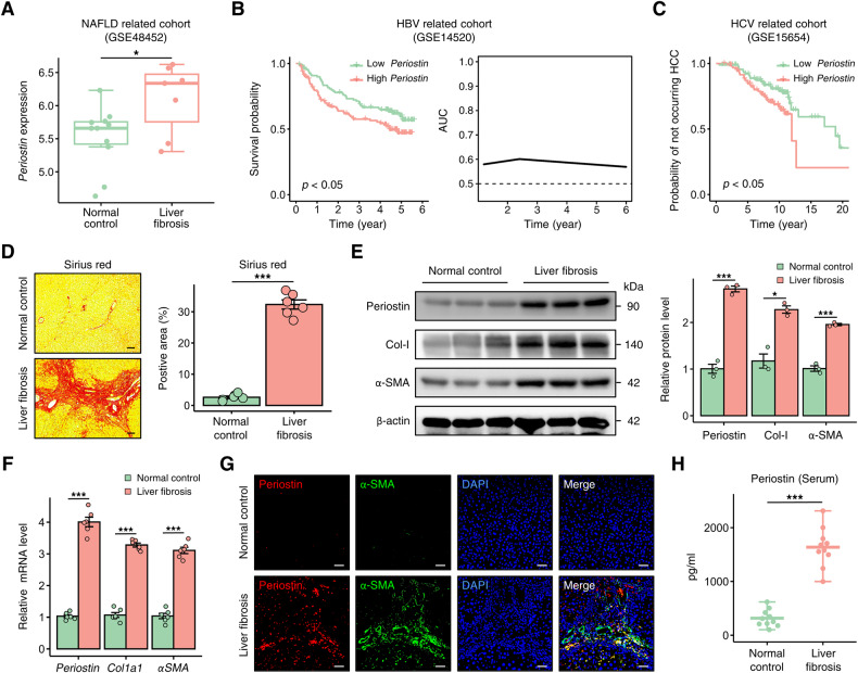
Fig. 4. Periostin is elevated in liver fibrosis patients.
A Periostin upregulation in the livers of patients with liver fibrosis in a NAFLD-related cohort. B Kaplan–Meier survival curves demonstrated that high Periostin levels in the liver were associated with a poorer overall prognosis compared to low Periostin levels in liver fibrosis patients infected with HBV (left). Predictive performance of liver Periostin levels in predicting the survival time of these patients with liver fibrosis (right). C HCV-infected liver fibrosis patients with high levels of Periostin in the liver exhibited an increased probability of developing HCC. D Sirius red staining was used to assess the extent of fibrosis in human samples from indicated groups. The data were quantified (n = 6 per group) (Scale bar: 100 μm). E, F Western blot and qPCR analyses revealed increased levels of Periostin, Col-I, and α-SMA in human liver samples with fibrosis. G Immunofluorescence staining showed that elevated Periostin is predominantly localized in α-SMA-positive regions of human fibrotic liver sections (Scale bar: 50 μm). H ELISA revealed the serum Periostin levels in normal control individuals and patients with liver fibrosis (n = 10 per group). All results are shown as mean ± SEM. *p < 0.05; ***p < 0.001. NAFLD non-alcoholic fatty liver disease, HBV hepatitis B virus; HCV hepatitis C virus; HCC hepatocellular carcinoma; Col-I Collagen-I.

