
Figure 1.
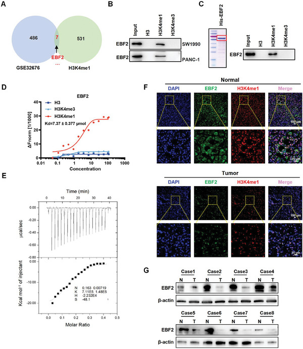
EBF2 is identified as an H3K4me1‐binding protein. A) Venn diagram showing the overlapping genes that were associated with H3K4me1 and significantly reduced in pancreatic cancer (GEO data, GSE32676). B) Peptide pull‐down assay to determine the interactions between H3, H3K4me1, or H3K4me3 peptides and EBF2 in vitro. C) Peptide pull‐down assay performed with purified His‐EBF2 fusion protein and H3, H3K4me1, or H3K4me3 peptides (right); Coomassie blue staining with SDS‐PAGE gel for the purified His‐EBF2 fusion protein expressed in E. coli (left). D) MST assay confirming the direct interactions between His‐EBF2 and H3, H3K4me1, or H3K4me3 peptides. Kd = 7.37 ± 0.377 µm. E) ITC binding curve of His‐EBF2 fusion protein with H3K4me1 peptide. Ka = 7.11 × 10⁵ ± 1.48 × 10⁵ mol−1. F) Immunofluorescence staining of EBF2 protein in PDAC and adjacent tissues. G) Western blot analysis for EBF2 protein level in PDAC and adjacent tissues.