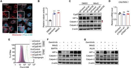
Figure 5.

TRAP1 inhibition induces mPTP opening, mitochondrial calcium discharge, and calpain‐1 activation. A) Visualization of cytoplasmic calcium. Fluo‐4 AM‐labeled MIO‐M1 cells were incubated with the TRAP1 inhibitors gamitrinib and MitoQ[ 32 , 41 ] for 6 h or thapsigargin for 30 min under hypoxia, and analyzed by confocal microscopy. Scale bars, 10 µm and 2 µm (inset). B) Calpain activation by TRAP1 inhibitors. MIO‐M1 cells were treated with a TRAP1 inhibitor, 3 µM gamitrinib or 0.5 µm MitoQ, for 6 h under hypoxia (n = 4). Enzyme activity was measured using a fluorogenic calpain substrate as described in the Experimental Section. C) Restored HIF1α expression upon CypD inhibition. MIO‐M1 cells were incubated with control or CypD‐targeting siRNAs, treated with the TRAP1 inhibitor MitoQ for 6 h under hypoxia, harvested, and analyzed by western blotting. Black and red arrows indicate pro and autolyzed forms of calpain‐1, respectively. D) Calpain‐1 mRNA expression upon TRAP1 depletion. MIO‐M1 cells were incubated with TRAP1‐targeting siRNAs for 48 h, exposed to hypoxia for 6 h, harvested, and analyzed by qPCR (n = 4). E) Modestly elevated cytosolic calcium by TRAP1 inhibition. After siRNA knockdown of CypD, Fluo‐4 AM‐labeled MIO‐M1 cells were incubated under hypoxic conditions with MitoQ for 6 h. Cells were then analyzed by flow cytometry to detect cytoplasmic calcium. F) Inhibition of HIF1α degradation by calcium chelation. MIO‐M1 cells were incubated with TRAP1 inhibitors, 3 µm gamitrinib, and 0.5 µM MitoQ, and a cell‐permeable calcium chelator, BAPTA (10 µM), for 6 h as indicated under hypoxia and analyzed by western blotting. G) Blocked HIF1α degradation by calpain inhibition. MIO‐M1 cells under hypoxia were incubated with 3 µm gamitrinib, 0.5 µm MitoQ, and 10 µm ALLN (calpain inhibitor) as indicated for 6 h and analyzed by western blotting. Data information: Data are expressed as mean ± SEM. Student t‐test, *** P < 0.001; ns, not significant.
