Figure 6.

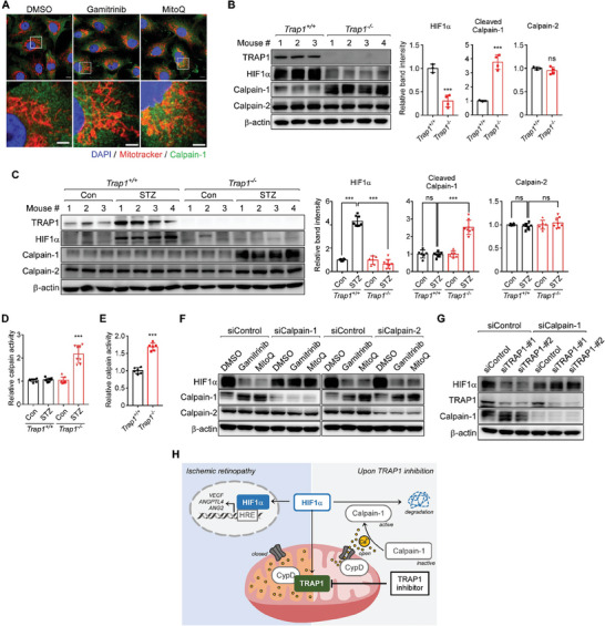
TRAP1 inhibition triggers calcium/calpain‐1‐dependent HIF1α degradation. A) Staining of mitochondria and calpain‐1. MitoTracker‐labeled MIO‐M1 cells were exposed to 3 µm gamitrinib or 0.5 µm MitoQ for 6 h under hypoxia and analyzed by immunocytochemistry with an anti‐calpain‐1 antibody. Scale bars, 10 µm (top) and 5 µm (bottom). B) Calpain autolysis in OIR mouse retinas. Left. Retinas collected from Trap1 +/+ (n = 3 mice) and Trap1 −/− (n = 4 mice) OIR mice were analyzed by western blotting. Right. Protein band intensities of HIF1α, cleaved calpain‐1, and calpain‐2 were normalized to those of β‐actin and compared. C) Calpain autolysis in STZ mouse retinas. Left. Retinal samples collected from STZ (n = 4 mice) and age‐matched control (n = 3 mice) mice with the Trap1 +/+ or Trap1 −/− genotype were analyzed by western blotting. Right. Protein band intensities were analyzed as in (B). D,E) Calpain activity in mouse retinas. Calpain enzyme activities were analyzed in retinas collected from Trap1 +/+ and Trap1 −/− STZ (D, n = 8 mice/group) and OIR (E, n = 6 mice/group) mice and compared. F) Depletion of calpains by siRNAs. Calpain‐1‐ and calpain‐2‐targeting siRNA‐treated MIO‐M1 cells were incubated with 3 µm gamitrinib or 0.5 µm MitoQ for 6 h under hypoxia and analyzed by western blotting. G) Depletion of calpain‐1 and TRAP1 by siRNAs. MIO‐M1 cells treated with siRNAs as indicated were exposed to hypoxia for 6 h and analyzed by western blotting. H) HIF1α degradation following TRAP1 inhibition. TRAP1 inhibition caused mild mitochondrial calcium discharge into the cytoplasm by opening CypD‐regulated mPTPs. Subsequent activation of calpain‐1 proteolytically degrades HIF1α. Data information: Data are expressed as mean ± SEM. Student t‐test, *** P < 0.001; ns, not significant.
