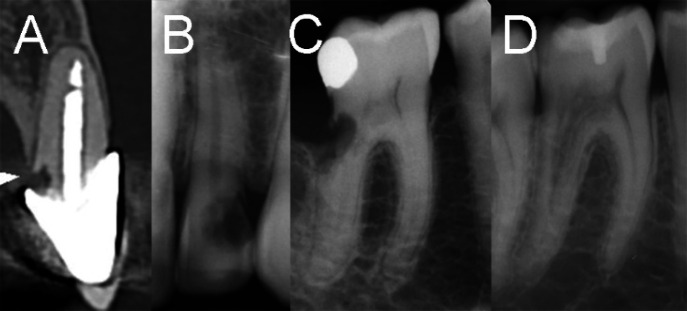
Figure 2.
Radiographic Presentation of various ICRR Cases; A) Class 1 exhibits a small lesion near the cervical area with limited penetration into the dentine; B) In Class 2, a well-defined ICRR lesion is observed close to the coronal pulp chamber; C) Class 3 signifies a deeper invasion, involving the coronal dentine and extending into the coronal third of the root; D) Finally, Class 4 indicates a large ICRR process that extends beyond the coronal third of the root

